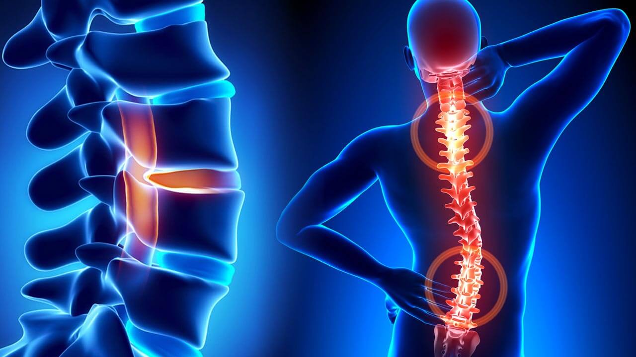
Curvature of The Spine
‘’Scoliosis’’, which is the name given to spinal curvature, is a condition that is typically diagnosed during the growth period. Scoliosis, which develops as a result of the spine curving or twisting to the right or left due to various reasons, can seriously affect a person’s life if left untreated, beginning at a young age.
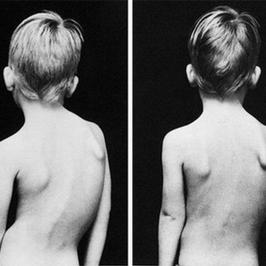
Scoliosis, which has a prevalence rate that varies between 0.2% and 6%, is the oldest known spinal deformity. While it can develop due to various causes such as trauma and congenital developmental disorders, the cause of 80% of skolyoz cases is unknown. Generally, at the beginning of the growth period, symptoms such as asymmetry of the shoulders, a protrusion in a part of the back, and the hips not being at the same level are noticed by the parents.
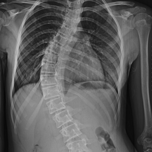
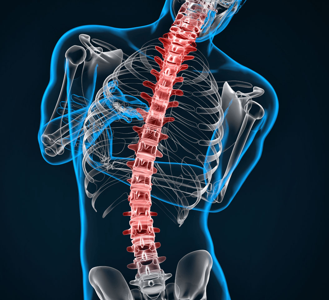
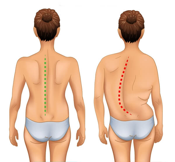
Skoliosis, also known as curvature of the spine, is when the spine bends to the side at an angle greater than 10 degrees. In a normal and healthy spine, the vertebrae run in a straight line from top to bottom, from the neck, through the back, and down to the lower back. However, in scoliosis, the vertebrae shift to the right or left and also rotate around their own axes. Therefore, it is defined as a three-dimensional deformity (shape abnormality).
Scoliosis can also cause shifts in the hips, ribcage, and shoulder blades, leading to postural and visual abnormalities. In children during their growth phase, this condition can cause abnormal loading on developing and growing spine and resulting in deformities in the vertebrae.
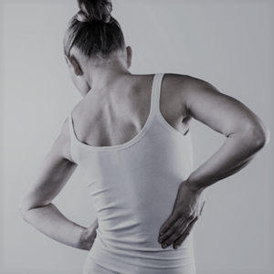
This condition is more common in boys in the preschool period and 3-5 times more common in girls during adolescence, depending on the growth rate. Scoliosis, which does not cause strong complaints in the patient in the early stages, is mostly detected by chance during school screenings or X-ray imaging for any reason. In addition, the child’s body appearance disorder is one of the most important reasons why families seek medical attention. Asymmetry in the shoulders, shoulder blades, breast level, and waist curves are the most noticeable findings. This can be accompanied by back and waist pain. As the degree of curvature increases, respiratory distress may also occur.
SYMPTOMS
- If there is asymmetry in the person’s back or waist,
- If one shoulder is higher than the other,
- If one shoulder blade is more protruding or prominent than the other when viewed from the back,
- If one leg appears longer than the other,
- If the trunk or chest is tilted to one side,
- If when the person leans forward, the ribs on one side of the body remain higher than the other side,
- If clothes constantly hang asymmetrically on the person, there may be a spinal deformity and suspicion should be raised.
ABOUT SCOLIOSIS
Scoliosis may not show any symptoms in the early stages. Even if scoliosis symptoms appear, they often do not cause much discomfort, so people may not take action. Complaint ıf any bile this is very low düzeydedir. It is usually detected by chance on X-rays taken for any reason or through school screenings. The first sign that leads families to consult a doctor is usually a visual distortion. The first noticeable sign in idiopathic scoliosis, which has an unknown cause, is one shoulder being higher than the other. Asymmetry in the shoulders, shoulder blades, waist, or torso is the most noticeable visual distortion. Back and lower back pain is present in 40% of cases. Curves above 50 degrees can cause respiratory distress.
The natural course of scoliosis may not always remain the same. Spinal curvature can progress, remain stable, or rarely improve. An increase of 5 degrees or more in spinal curvature in two or more consecutive exams with a curve of 20 degrees or more, or an increase of 10 degrees or more in curves below 20 degrees, is considered progression. Double curves, curves in the thoracic region, female gender, and larger curve magnitude at the time of diagnosis, as well as curves diagnosed before the age of 10, are prone to progression. The progression rate is quite low for curves below 30 degrees. The degrees of scoliosis are listed as follows;
Chronological Classification
- Infancy: Between 0-2 years
- Juvenile period: Between 3-9 years
- Adolescent period: Between 10-17 years
- Adulthood Period: 18 years of age and older
Classification by Placement
When examining the anatomy of scoliosis, it can be classified into cervical vertebrae, cervical and upper thoracic region, regional thoracic vertebrae, lumbar and lower thoracic vertebrae, and regional lumbar vertebrae.
Angular Classification
Imaging methods are used to grade the angular degree of scoliosis. After the imaging method, the curvature of the spine is diagnosed in terms of angle. This method is particularly useful in determining the need for surgical intervention in scoliosis.
Angle below 10 degrees: Called “spinal asymmetry” in medical terms, does not have any impact on their health. An angle above 10 degrees is necessary for treatment of the curvature. Low degree curvatures should still be monitored through periodic check-ups to ensure they do not pose a risk of developing scoliosis in the future. Here, the important thing is to determine whether scoliosis is progressing or not.
Angles between 20 and 40 degrees: Curvatures between 20 and 40 degrees are mostly seen during adolescence. This degree, which is considered as moderate scoliosis, is usually treated effectively with exercise, physical therapy and braces.
Angles of 40 degrees: Scoliosis curvatures of 40 degrees have largely completed their growth and progression. For surgical intervention to be performed, the back curvature should be over 45-50 degrees and the curvature in the lumbar region should be 40 degrees.
In 80% of scoliosis patients, the cause of the curvature cannot be determined. However, when looking at structural abnormalities that cause scoliosis, it can be said that congenital structural abnormalities, nerve and muscle diseases (cerebral palsy, syringomyelia, polio, muscular diseases, etc.), spinal tumors, trauma, spinal infections, and metabolic diseases can cause scoliosis. In addition, postural disorders and differences in leg length are also among the causes of scoliosis. The causes of scoliosis can be briefly explained as follows;
- Congenital scoliosis caused by structural abnormalities in the spinal bone structure
- Infantile and juvenile scoliosis that begins in early childhood
- Scoliosis caused by neuromuscular reasons such as muscular dystrophy
- Scoliosis caused by connective tissue diseases such as Marfan syndrome and Ehlers-Danlos syndrome
- Scoliosis caused by polio inflammatory diseases and traumas
- Scoliosis caused by leg length discrepancy and hip and knee joint problems.
The diagnosis of scoliosis can be determined through a child’s examination. When viewed from the front against a bare spine, asymmetry in the midline can be noticed. When the child bends forward, a tilt to one side and a rib hump on the side where the curve is present become apparent. This appearance is called a rib hump or hunchback. It may be difficult to notice this image in some cases of “balanced scoliosis”.
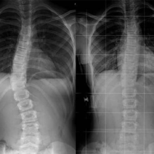
The first step in diagnosing scoliosis is to take an X-ray. The aim is to confirm the curvature of the spine, determine its size and location, and identify any hereditary bone disorders associated with it. X-rays should be taken every six months to monitor scoliosis. On the other hand, other imaging tests such as bone scintigraphy, computed tomography (CT), or magnetic resonance imaging (MRI) may be performed for patients with neurological disorders or those who will undergo surgery.
Diagnosis can easily be confirmed with direct radiographs taken for suspicion of scoliosis. Rarely, an MRI may be necessary. As radiological tests are frequently used for scoliosis monitoring and diagnosis, great care should be taken to protect the ovaries and breasts of these children with lead shields.
Scoliosis curvatures are defined as major and minor curvatures. The location where the curvature is most pronounced, i.e. the vertebrae that rotate the most from the vertical axis and are farthest from the midline, is called the apex. The type of scoliosis is named according to the level of the spine where the apex is located. If the apex is in the neck region, it is called cervical scoliosis, if it is in the back region, it is called thoracic scoliosis, and if it is in the lower back region, it is called lumbar scoliosis. Sometimes it can be seen in multiple areas at the same time:
For example, when it occurs both in the back and in the waist, it is defined as thoracolumbar scoliosis. It is more common in the back (thoracic) area.
The form and degree of scoliosis are determined in the taken X-rays. The most commonly used method for this is the Cobb angle. Scoliosis is monitored by the Cobb angle and age of growth, and appropriate treatment methods are decided. The Cobb angle is measured by drawing lines from the upper limit of where the bending begins to the lower limit of where the bending ends on the spine. The angle between these lines drawn to these limits (i.e., the axis of the spine where the curvature begins and the axis of the spine where the curvature ends) is examined.
Sometimes scoliosis can spontaneously regress. It is not possible to predict how scoliosis that occurs at the beginning of the growth period will progress. Some recent studies have shown that there may be progression in children who carry certain genetic traits. Important monitoring criteria are used to determine the treatment of scoliosis. In some cases, progression is common, and the success rate of treatments is lower. These are as follows;
- High degree of curvature at the time of initial diagnosis
- Double curvature in both the back and waist,
- Neuromuscular scoliosis
- Serious contracture and muscle shortening
The decision on how to treat is made considering the risk of progression of spinal curvature. The accepted treatment methods for scoliosis are as follows:
- Monitoring and continuous development
- Brace applications
- Scoliosis exercises and special rehabilitation applications
- Surgery
In children who have not yet started growing and have a Cobb angle below 15 degrees, specialist follow-up is usually recommended. For those with a Cobb angle between 15-20 degrees, special scoliosis exercises and rehabilitation programs should be continued. Intensive scoliosis rehabilitation programs should be implemented for children with a Cobb angle of over 25 degrees.
During the adolescent period when the first signs of growth appear, such as hair growth, voice changes, increased height in boys, the beginning of breast development or the onset of menstruation in girls, great care should be taken and children should be treated. Since the curvature rate and risk are higher in these children, the progression or progression risk should be calculated rather than the degree of Cobb angle, and treatments should be planned accordingly. Children with a high risk of progression should use a brace in addition to physiotherapy and rehabilitation treatments. Brace therapy should be continued for 16 to 23 hours a day depending on the growth status and degree of curvature until growth is completed.
If brace therapy is unsuccessful and the Cobb angle is above 50 degrees in individuals with a high risk of progression, surgical treatment may be applied. In scoliosis surgery, the spine is taken in the midline with plates and screws, and these metals sometimes remain permanently in these children’s bodies. Late complications of surgery should also be considered.


